Fig. 4.
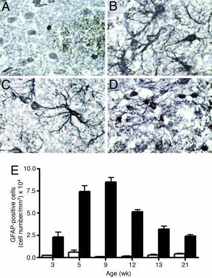
Reactive astrogliosis in MUT mice. GFAP immunohistochemistry in WT (A) and MUT (B–D) at varying ages (B, 5 wk; C, 9 wk; D, 21 wk). (E) The number of GFAP-positive cells was quantified at different ages. There is significant difference (P < 0.05) between the number of cells in WT (white bars) and MUT (black bars) at all ages.

