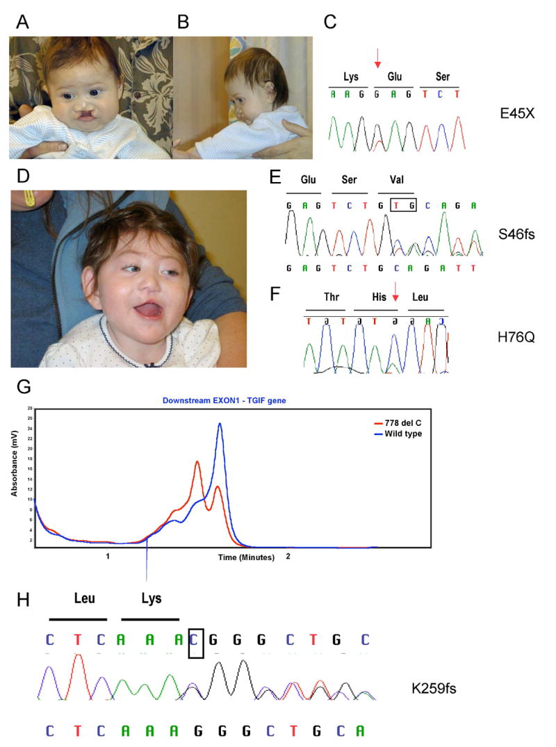
Fig. 1.

Facial photographs of the first proband (A and B) with the pE45X mutatiom (C). Patient two (D) carries two different variations the S46fs (E) and H76Q (F). Red arrows indicate the missense changes and the deleted bases are boxed. The expected chromatogram from the normal allele is above and the frameshifted allele is below. A representative dHPLC chromatogram from patient three (G) shows heterozygosity for the normal allele (blue) and the variant allele (red). In panel H, the deleted base is boxed and the expected sequence from the normal allele is above and the frameshifted allele is below the chromatogram.
