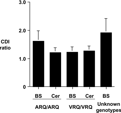
Figure 4. Low CDI values for scrapie-negative ovine brain tissue.
Scrapie-free brain homogenates were prepared from brain stem (BS) and cerebellum (Cer) of homozygous ARQ and VRQ sheep, and from brain stem of Bio-Rad TeSeE-negative sheep of unknown PrP genotypes, as described in the Materials and methods section. Brain homogenates, treated with or without 6 M GdnHCl, were subjected to CDI using mAb FH11 as capture and mAb V24 as detector. The results shown are CDI ratios (denatured/native fluorescence cps)±S.D. for scrapie-free brain stem and cerebellum brain tissue.

