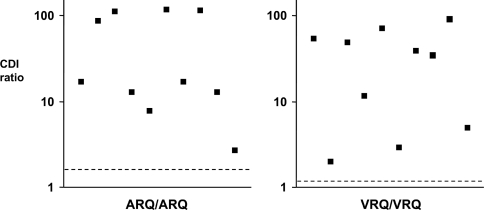
Figure 6. CDI ratios for homozygous ARQ and VRQ scrapie-infected brain stems from natural cases of typical scrapie.
Homogenates were prepared from brain stem of known cases of homozygous ARQ (left-hand panel) and VRQ (right-hand panel) scrapie-infected sheep as described in the Materials and methods section. PrPSc was isolated by sarkosyl extraction and subjected to CDI using anti-PrP mAb FH11 as capture and anti-PrP mAb V24 as detector. The results shown are individual CDI ratios (denatured/native fluorescence cps) for each tissue sample. The broken line is the mean CDI value for scrapie-free tissue of the same genotype (n=10 for each genotype).

