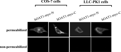
Figure 2. Immunofluorescent localization of Myc-tagged hOAT1 in COS-7 cells and LLC-PK1 cells.
Cells transfected with cDNAs encoding hOAT1–Myc-N or hOAT1–Myc-C were visualized with indirect immunofluorescent labelling. Permeabilized cells were generated by treatment with 0.1% Triton X-100. Proteins were detected with anti-Myc antibody as primary antibody and FITC-conjugated goat anti-mouse IgG as secondary antibody. Specific immunostaining appears as green fluorescence.

