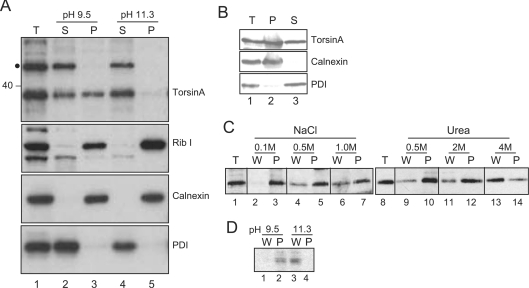
Figure 3. TorsinA is peripherally associated with the ER membrane.
(A) HeLa cells were mechanically cracked and extracted twice with CAPSO buffer (pH 9.5) or sodium carbonate (pH 11.3). Membrane-associated material was isolated by centrifugation, and proteins in the supernatant precipitated with TCA. Samples of the total lysate (T), supernatant (S) and membrane pellet (P) were analysed by Western blotting with affinity purified anti-torsinA, or antibodies to ribophorin I (Rib I), calnexin or PDI. ● shows an approx. 50 kDa protein that cross-reacts with the torsinA antisera. (B) HeLa cells were permeabilized with 0.2% saponin and isolated by centrifugation. Samples of total cells (T), supernatant (S) and cell pellet (P) were analysed by Western blotting with anti-torsinA. (C and D) Saponin-permeabilized cells were washed with buffer containing NaCl, urea, CAPSO buffer (pH 9.5) or sodium carbonate (pH 11.3). Cells were reisolated by centrifugation. Samples of the total cells (T), wash (W) and cell pellet (P) were analysed as in (B).

