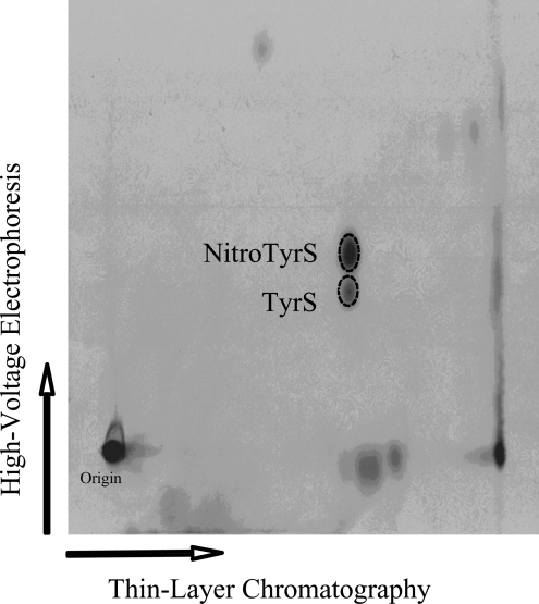
Figure 5. Identification of free nitrotyrosine O-[35S]sulfate and tyrosine O-[35S]sulfate generated and released by HepG2 human hepatoma cells.
Autoradiograph taken from the TLC plate used for the two-dimensional thin-layer analysis of the labelling medium sample. Confluent HepG2 cells were incubated for 18 h in the labelling media containing 2.5 mM SIN-1. The broken-line circles correspond to the positions of synthetic nitrotyrosine O-sulfate (upper) and tyrosine O-sulfate (lower) as revealed by ninhydrin staining.

