
Fig. 7.
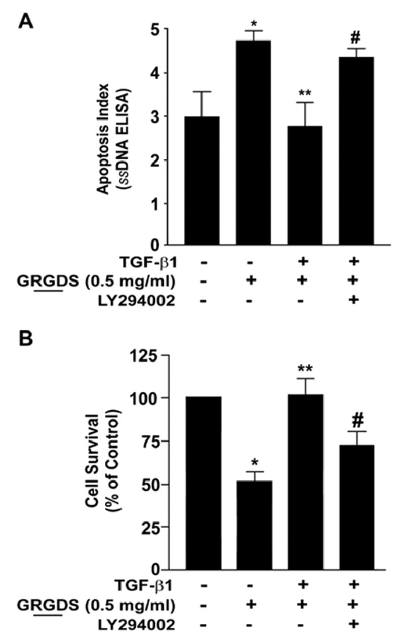
Myofibroblast viability and anoikis-resistance is dependent on PI3K-AKT. (A) Quiescent IMR-90 fibroblasts were stimulated with/without TGF-β1 in the presence/absence of LY294002 (10 μM) for 16 h prior to treatment with soluble RGD-containing peptides (GRGDS, 0.5 mg/ml) for 24 h and assessed for viability as described in “Materials and methods”. Values are mean±SEM; n=3 and experiments was repeated 3 times with similar results. * p<0.01 compared to untreated controls. ** p<0.001 compared to GRGDS 0.5 mg/ml alone. #=not significant compared to 0.5 mg/ml GRGDS alone. (B) IMR-90 cells were cultured in a 96 well plate, growth-arrested and treated±TGF-β1 with/without the PI3K inhibitor LY294002 (10 μM) for 16 h prior to the addition of soluble RGD containing fibronectin peptide (GRGDS 0.5 mg/ml) for 24 h. Apoptosis was assessed with ELISA for ssDNA. Values are mean±SEM; n=4 per condition, and the experiment was repeated three times with similar results. * p<0.05 compared to untreated control. ** p<0.05 compared to 0.5 mg/ml GRGDS treatment alone. #=not significant compared to 0.5 mg/ml GRGDS alone.