
FIGURE 4. Phagocytosis of zymosan by group V sPLA2-null macrophages.
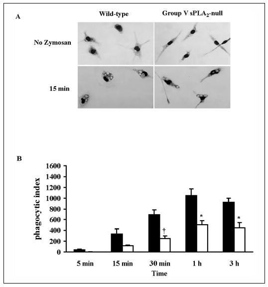
A, light microscopy image of peritoneal macrophages derived from group V sPLA2-null mice (BALB/c N3) and wild-type littermates stained with Diff-Quick before and 15 min after the addition of zymosan (10 particles/cell). B, peritoneal macrophages from group V sPLA2- null mice (open bars) and from their wild-type littermate controls (filled bars) bred to a BALB/c background for three generations were incubated with 10 particles of zymosan/cell for 5 min to 3 h and stained with Diffquick. The phagocytic index was calculated as described under “Experimental Procedures. ” †, p < 0.02; *, p < 0.05 using Student’s paired t test.