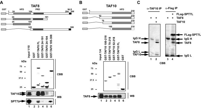
Figure 2. TAF8 interacts with TAF10 through its HF domain, while it interacts with SPT7L through its C-terminal region.
GST-TAF8 (A) and GST-TAF10 (B) full length (FL) or the corresponding deletion mutants were immobilised on glutathion-sepharose beads (as indicated), washed and the bound GST-fusion proteins were separated by SDS-PAGE and visualized by Coomassie brilliant blue staining (CBB). (*) labels premature termination products. Equal amounts of the indicated baculovirus infected Sf9 whole cell extracts (WCEs) were incubated with each resin, washed and the bound proteins analysed by western blot (WB). The positions of molecular weight markers are indicated in kDa. HFD: histone fold domain; PRD: proline rich domain; NLS: nuclear localization signal. (C) TAF8 was expressed alone (lane 1 and 3), coexpressed with TAF10 (lane 2) or with Flag-SPT7L (lane 4) in Sf9 cells using the baculovirus system. From these cells WCEs were prepared and proteins immunoprecipitated with the indicated antibodies. Bound proteins were separated by SDS-PAGE and visualized by Coomassie brilliant blue staining (CBB). The co-immunoprecipitation between TAF8 and TAF10 as well as TAF8 and SPT7L indicates strong stoichiometric interactions between these proteins.

