
Fig. 5.
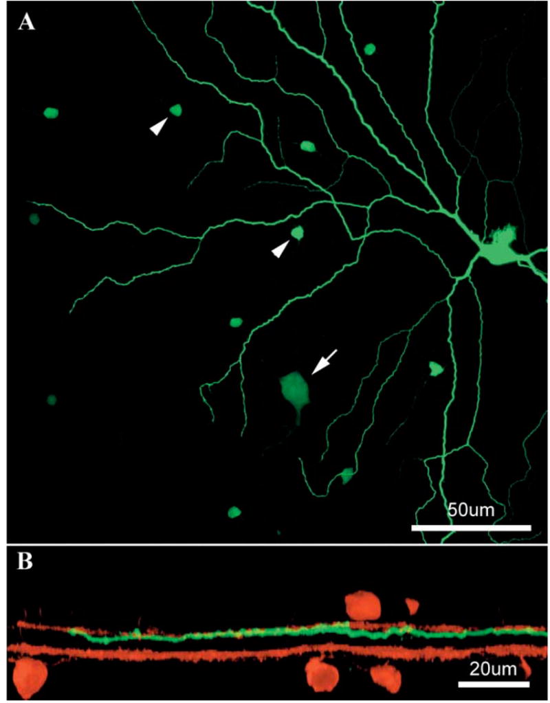
Double-label confocal images of an OFF α ganglion cell. A: Wholemount view shows part of the dendritic field of a Neurobiotin injected OFF α cell (green). The injected soma is marked by some dye leakage. This ganglion cell type is typically dye coupled to amacrine cells (arrowheads) and other OFF α cells (arrow). B: Z-axis reconstruction shows that the OFF α ganglion cells is stratified just below cholinergic a in the IPL. The cholinergic strata are labeled red.