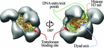
Fig. 4.
Model of nucleosome binding by RSC. The x-ray crystal structure of the nucleosome (23) (PDB ID code 1AOI) was manually fitted into the central cavity of RSC. The nucleosome is shown as a ribbon diagram within a translucent surface representation filtered to 10 Å. The DNA is represented in gold, and the protein is represented in orange. Back (Left) and front (Right) views of the complex are shown. The entry/exit points of the nucleosomal DNA are indicated with green arrows, the dyad axis (blue cylinder) is indicated with a blue arrow, the histone H3 tail visible in the crystal structure is indicated with an orange arrow, and the binding site for the translocase domain is shown on the DNA with maroon arrows.

