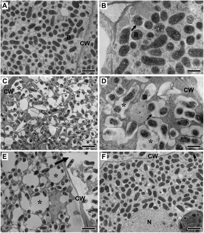
Figure 4.
Ultrastructure of infected cells of wild-type and ign1 nodules. A and B, Wild-type nodules at 10 (A) and 13 dpi (B). C to E, ign1 nodule infected cells at 8 (C), 10 (D), and 13 dpi (E). Irregularly shaped, enlarged symbiosomes are indicated by asterisks. An arrow in E indicates a collapsed infected cell. F, Infected cells of 10 dpi nodules formed by a ΔnifH mutant strain of M. loti. CW, Cell wall; B, bacteroid; N, nucleus. PBM is indicated by arrows in B and D. Bars indicate 2 μm (A, C, E, and F) and 1 μm (B and D).

