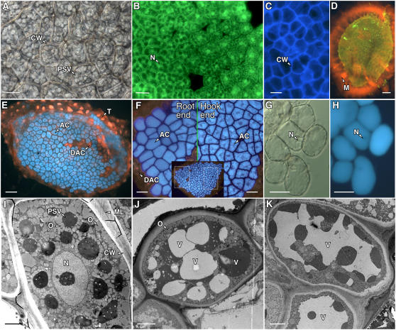
Figure 6.
Cells in the Arabidopsis endosperm are typical aleurone cells. Differential interference contrast (A and G), fluorescence (B–F, H), and electron microscopy (I–K) images of Arabidopsis aleurone layers. A, Region of an aleurone layer showing aleurone cells surrounded by a thick cell wall (CW) and containing numerous PSVs. B, Aleurone layer labeled with the vital fluorescent probe fluorescein diacetate showing green fluorescence from the cytosol and nucleus (N) of live cells. C, Cellulose in the Arabidopsis aleurone cell wall labeled with calcofluor white M2R. D, An extensive mucilage layer (M) that was labeled with orange fluorescent Congo red was retained by the testa cells associated with the isolated aleurone layer. E, Aleurone cells (AC) incubated with monochlorobimane accumulated blue fluorescent bimane in their PSV. The testa and nuclei in dead aleurone cells (DAC) in E fluoresced red after labeling with propidium iodide. F, Cells in Arabidopsis aleurone layers closest to the root became less angular in shape sooner than cells near the hypocotyl hook. Shown are higher magnification images that correspond to the boxed regions of the single aleurone layer shown in the inset. G, High magnification image of individual aleurone cells close to the site of root emergence. Cell walls had begun to separate, and individual cells had become nearly spherical. H, Cells in G are still alive, as indicated by the fluorescence from the vacuole and lack of propidium iodide fluorescence from the nucleus (N). I, Electron micrograph of an aleurone cell 1 h after imbibition showing the numerous PSVs and oleosomes (O). The aleurone cell wall and middle lamella (ML) are also indicated. J and K, Aleurone cells 24 h after imbibition. Note the enlargement of the vacuoles (V) and reduction in the number of oleosomes compared with those in I. The vacuoles in J are heterogeneous with regard to electron density. Scale bars are 2 μm in I to K, 10 μm in A, G, and H, 20 μm in B, C, and F, and 60 μm in D and E.

