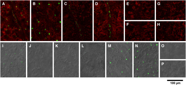
Figure 2.
Confocal Microscopic Observation of cry2-GFP Nuclear Accumulation (Guo et al., 1999) in Mesophyll, Vascular Bundles, and Epidermis of Cotyledons.
Seedlings were grown for 10 d under LD. Green fluorescence from GFP and red fluorescence from chlorophyll were overlaid electronically. In addition, differential interference contrast images were overlaid for epidermis. Seedlings of pCRY-C2G-16 ([A] and [I]), pCAB-C2G-6 ([B] and [J]), pSUC-C2G-2 ([C] and [K]), pSultr-C2G-10 ([D] and [L]), pML-C2G-6 ([E] and [M]), pCER-C2G-8 ([F] and [N]), pUFO-C2G-13 ([G] and [O]), and pAt3g-C2G-7 ([H] and [P]) are shown. Bar = 100 μm.
(A) to (H) cry2-GFP fluorescence in mesophyll/vascular bundles.
(I) to (P) cry2-GFP fluorescence in epidermis.

