
Figure 1.
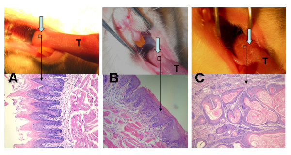
Both gross and microscopic changes in SD rats' tongues mucosas. A. Normal mucosa of tongue base at 6th week (gross observation and HE ×200); B. Epithelial dysplasia of tongue base at 24th week (gross observation and HE ×200); C. Squamous cell carcinoma of tongue base at 36th week (gross observation and HE ×200). T represents tongue.