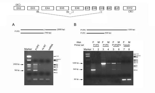
Figure 2.
mfl encodes a novel alternatively-spliced mRNA. At the top, a schematic drawing of the splicing patterns generating mfl mRNAs; the position and structure of the primers utilised in RT-PCR assays are indicated. HindIII-digested λ DNA was used as marker; base pairs are indicated on the right. (A) RT-PCR performed with poly(A)+RNA isolated from adult females and an exon 2–12 primer pair (P1/P2). The 2000 bp fragment is specific for the 2.2 kb mRNA, while the 740 bp fragment derives from the 1.0 kb alternatively-spliced subform. (B) RT-PCR was performed with poly(A)+RNA isolated from male (M) and female (F) adult flies. Female or male samples were amplified using the same forward primer (P1, derived from exon 2) in combination with a reverse primer spanning the alternative 3–9 exon junction (P3; lanes 1–2) or the canonical 5–6 exon junction (P4; lanes 3–4); in lanes 5–6, the three primers were added to the same reaction. The 1200 bp fragment represents all three transcripts generated by the canonical splicing pattern, while the 540 bp fragment derives specifically from the 1.0 kb mRNA. In lanes 7–8, a 90 bp fragment representating the αTub84B transcript was amplified as internal control of the quantity of RNA.

