Figure 2.
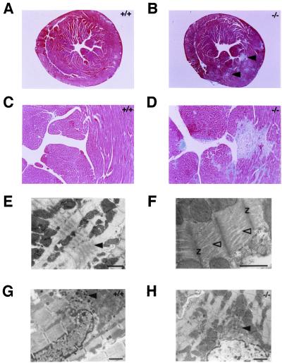
Light and electron microscopic examinations of hearts from Nppb+/+ and Nppb−/− mice. (A and B) Masson's trichrome staining of transverse sections of ventricles from Nppb+/+ (A) and Nppb−/− (B) mice at the level of papillary muscles. Focal fibrotic lesions are indicated by arrowheads. (C and D) Higher magnification (×25) of subendocardial regions of ventricles from Nppb+/+ (C) and Nppb−/− (D) mice. (E and F) Electron microscopy of left ventricular free wall from Nppb−/− mice. Supercontracted sarcomeres and disorganized myofibrils are indicated by filled and open arrowheads, respectively. Z, Z-bands in sarcomeres. (G and H) Electron microscopy of atria from Nppb+/+ (G) and Nppb−/− (H) mice. Atrial granules are indicated by arrowheads. (Scale bars represent 1 μm in E–H.)

