
Figure 1. Expression of HSV-1 LAT increases the percentage of substance P-immunoreactive trigeminal neurons.
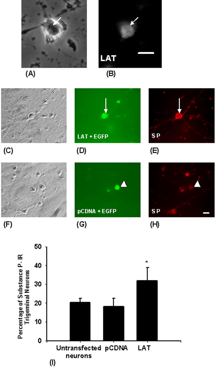
In order to Express LAT in trigeminal neurons, cultured trigeminal neurons were cotransfected with plasmids expressing LAT and EGFP as a marker. The upper panel shows a trigeminal neuron transfected with LAT containing plasmid (A), and LAT was detected in the nucleus of the neuron (indicated by arrows) by in situ hybridization (B). Two days after transfection, the cells were fixed and immunostained for substance P. The middle panel shows a trigeminal neuron (indicated by arrows) cotransfected with LAT and EGFP (D) and this neuron was substance P-immunoreactive (E). The lower panel shows a trigeminal neuron (indicated by arrowheads) cotransfected with pCDNA3.1 and EGFP (G) and that neuron showed only background staining (not immunoreactive to substance P) (H). LAT expression resulted in a two-thirds increase in the percentage of substance P immunoreactive trigeminal neurons (I). Bars are means ± S.E.M. (∗) indicates p < 0.05. Scale bar = 40μM.