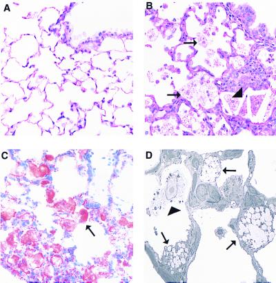
Figure 5.
Pulmonary lesions in Abc1−/− mice. A contains a section of normal lung from a 12-mo-old +/+ mouse with a normal terminal bronchiole and alveoli lined predominantly with flattened type I pneumocytes (hematoxylin/eosin stain, ×400). B demonstrates an early lesion from a 12-mo-old −/−mouse including numerous foamy cells (arrows) and cholesterol clefts (arrowheads). Alveolar septae are focally expanded by mild type II pneumocyte hypertrophy, macrophages, and aggregates of lymphocytes and plasma cells. An Oil red O-stained section of lung from a 7-mo-old −/− mouse demonstrates lipid accumulation within alveolar cells (arrow, C). By electron microscopy, these cells are identified primarily as type II pneumocytes (D, arrows) and intraalveolar macrophages (arrowhead, ×3,500).

