
Figure 1.
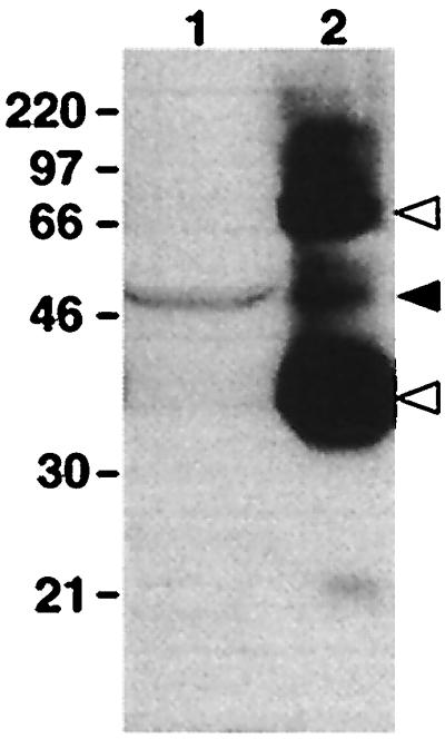
Western blot of protein extracts from dermal melanophores and whole eye probed with an antiserum raised against bovine rhodopsin. Indicated molecular masses are in kDa. Lanes: 1, total protein from cultured melanophores; 2, 1% of total protein from a whole early postmetamorphic adult eye. A 50-kDa immunoreactive band is present in both lanes (solid arrowhead). The 35- and 70-kDa bands in lane 2 are monomeric and dimeric rhodopsin, respectively (open arrowheads).