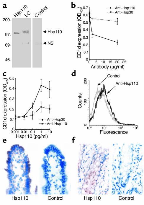
Figure 6.
Role of Hsp110 in induction of epithelial CD1d. (a) Western blot analysis of LC for Hsp110. LC or purified recombinant Hsp110 (0.3 μg) was resolved by SDS-PAGE under nonreducing conditions, and blots were probed with polyclonal rabbit anti-Hsp110. Also shown is the control with primary antibody omitted from the reaction mixture. A nonspecific band (NS) of approximately 70 kDa was observed in both LC and control samples. (b) Cell surface ELISA of T84 monolayers following incubation with 6 μg/ml LC in the presence of indicated concentrations of anti-Hsp110 or control anti-Hsp30. (c) Cell surface ELISA of T84 monolayers following incubation with indicated concentrations of purified recombinant Hsp110 in the presence of anti-Hsp110 or control anti-Hsp30. OD values generated by ELISA were blanked against isotype-matched control antibodies. (d) Flow cytometric analysis of Hsp110 on native small intestinal enterocytes (black line) compared with equivalent concentrations of an isotype-matched control (gray line). (e and f) Immunohistochemical localization of Hsp110 in normal small intestinal tissue (e) and in normal colon (f). Positive staining with anti-Hsp110 but not with control Ig (Control) is seen in epithelial cells and diffusely in the lamina propria. Magnification, ×400.

