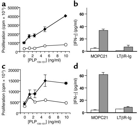
Figure 5.
LTβR-Ig treatment impairs T cell recall responses to PLP139–151. (a) On day 7 after immunization with PLP139–151 in SJL mice, pooled LN cells from control huIgG (diamonds) and LTβR-Ig–treated (circles) mice were isolated, stimulated in vitro with PLP139–151, and proliferation was measured by 3H-thymidine uptake. (b) Supernatants from cultures in a (0 and 10 μg/ml PLP139–151, white and gray bars, respectively) were collected at 72 hours and measured by ELISA for IFN-γ content. (c) Proliferation data as in a for splenocytes isolated on day 59 after immunization. (d) Supernatants from cultures in c (0 and 10 μg/ml PLP139–151) were collected at 72 hours and measured by ELISA for IFN-γ content. The experiment was also performed at day 10 and day 39 with similar results.

