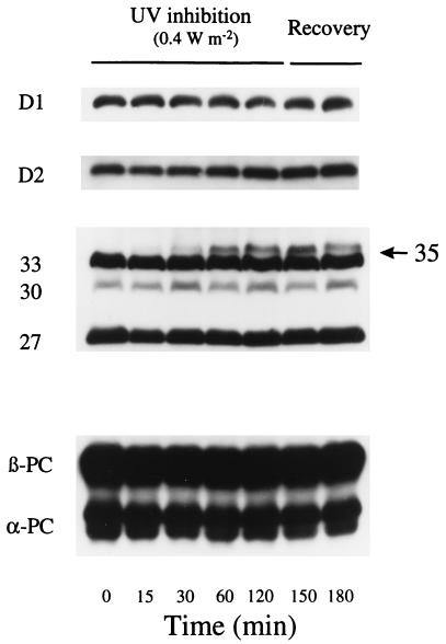
Figure 5.
D1, D2, and phycobilisome protein content during moderate UV-B exposure and recovery. Wild-type cells were supplemented with UV-B at 0.4 W/m2 for 2 h and then allowed to recover for 1 h without UV-B as described in Fig. 3. Total D1 (D1:1 and D1:2), D2, and phycobilisome were detected by using specific polyclonal antibodies. The figure shows results representative of three replicates.

