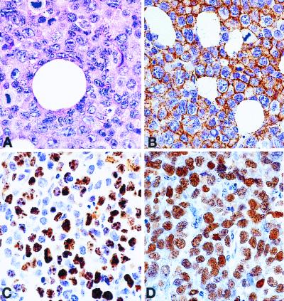
Figure 4.
Morphology and phenotype of the 20A cell line, xenotransplanted into a SCID mouse. (A) Hematoxylin-eosin stain showing large cell lymphoma with high mitotic rate. (B) Immunoperoxidase stain with antibody against human CD20 (B cell antigen) showing cell-membrane staining in all lymphoma cells. (C) Immunoperoxidase stain with an antibody against Ki-67 (cell proliferation-related antigen) showing nuclear staining in 50–80%. (D) In situ hybridization for EBV-encoded RNA1 (EBER1) showing nuclear positivity in all lymphoma cells.

