Figure 1.
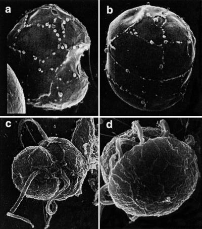
Scanning electron micrographs of P. piscicida (a–c) and the related dinoflagellate, Pfiesteria species B (d). (a) P. piscicida culture MMRCC98-1020BR01C5 (Florida DEP) stripped of the outer wall layer. Sutures between plates clearly delineate X, 1′,2′, 4′, 1" 5" s.a., SD, and s.s. plates. The cell is 6.1 μm long. (b) Anterior dorsal view of stripped, swollen cell from same culture, revealing plates Po, Cp, X, 2–4′, 1a, and 2–4" in the epitheca. The cell is 7.3 μm long. The entire plate formula for this genus is Po, Cp, X, 4′, 1a, 5", 6c, 4s, 5′′′, 0p, and 2′′′. In addition to number of plates, position and shape are also important; note the small triangular 1a plate (arrow). (c) Ventral view, P. piscicida culture NCSU 113-2. Suture lines delineate 1′ and adjacent plates. (d) Pfiesteria species B culture NCSU 7-28-T; note the “diamond” (quadrangular as opposed to triangular) cingular plate.

