Figure 1.
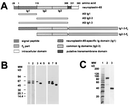
Characterization of neuroplastin antibodies and fusion proteins. (A) Map of np65 (np55 lacks the first Ig domain). The recombinant fragments to produce rabbit antisera AS Ig1, AS Ig2–3, and AS Ig1–3 are indicated below the map. The epitope for mAb SMgp65 is located within the Ig2–3 region. The Ig1–3-Fc and Ig2–3-Fc fusion proteins produced in 293 cells are also shown. (B) Specificity of antibodies. Western blots of Triton X-100-solubilized membrane proteins from rat brain (lanes 1–3 and 6), np65-transfected 293 cells (lanes 4 and 7), and np55-transfected 293 cells (lanes 5 and 8) were probed with the following antibodies: AS Ig2–3 (lane 1), AS Ig1–3 (lane 2), AS Ig1 (lanes 3–5), and mAb SMgp65 (lanes 6–8). Note, differential glycosylation of neuroplastins expressed by 293 cells results in size differences from neuroplastins of brain membrane extracts (23, 26). mAb SMgp65 recognizes two glycoforms of np65 in transfected 293 cells (lane 7, and ref. 23). (C) Purification of the Fc-fusion proteins. Affinity-purified fusion protein Ig1–3-Fc (lane 2) and human Fc alone (lane 3) were separated by SDS/PAGE and stained with Coomassie brilliant blue. Molecular weight markers are shown in lane 1.

