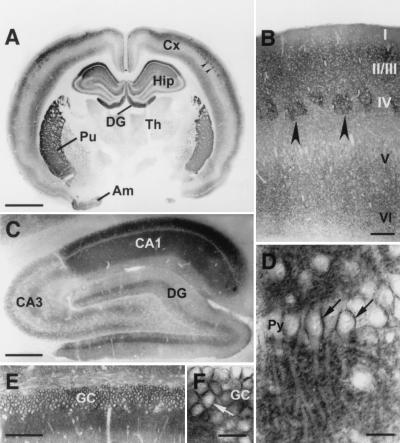
Figure 2.
Distribution of np65 in the forebrain detected with np65-specific antibodies (AS Ig1). (A) Frontal section of rat forebrain. Intense immunoreactivity is found in the cerebral cortex (Cx), the hippocampal formation (Hip), including dentate gyrus (DG), the putamen (Pu), and the amygdala (Am). Th, thalamus. (B) Frontal section of somatosensory cortex; layering is indicated. Arrowheads in A and B mark strongly stained barrel fields. (C) Sagittal section of the hippocampus. CA1, CA3, Ammon's horn regions 1 and 3. (D–F) Enlargements of the CA1 pyramidal cell layer (Py) and the granule cell layer (GC) of the DG indicate cell surface staining of these neurons (arrows). (Size bars: 2 mm in A; 200 μm in B; 500 μm in C; 25 μm in D and F; 100 μm in E.)

