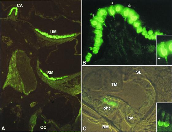
Figure 2.
KCNQ4 in the murine inner ear. (A) Immunofluorescence of an inner ear section at P21. KCNQ4 immunoreactivity is present in hair cells of the crista ampullaris (CA), utricular macula (UM), and saccular macula (SM), as well as in the OHC of the organ of Corti (OC). (B) Section of a crista ampullaris at P21. Cells are labeled at their basal and lateral membrane. Some of these cells (indicated by asterisks) can be unambiguously identified as type I hair cells because of the costaining of their characteristic ensheathing nerve calyx (arrowhead in Inset). Note that the staining of the calyx extends to the adjacent region of the nerve fiber. (C) Transverse section of the organ of Corti at P13 (transmitted light interference contrast). The three OHC (ohc) are labeled at their basal membrane, whereas the IHC (ihc) is not. A detailed view of two OHC is presented in the inset. BM, basilar membrane; SL, spiral limbus; and TM, tectorial membrane.

