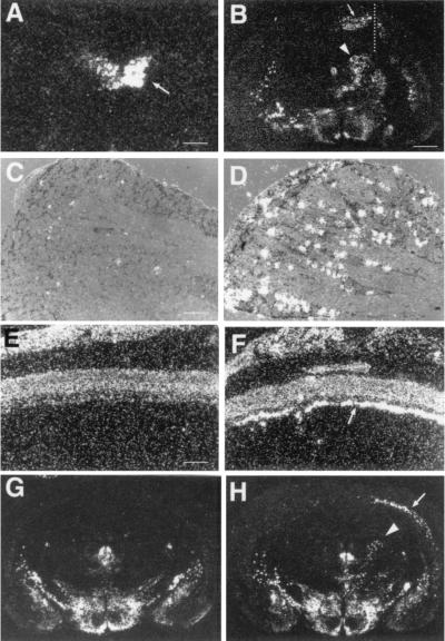
Figure 3.
DINE mRNA expression up-regulated by a variety of neuronal damage. (A) Hypoglossal nerve transection (arrow indicates hypoglossal nucleus of injured side). (B) Cortical and corpus callosum transection (the right cerebral cortex was cut by a knife sagittally; broken line). DINE mRNA is expressed in the cortical neurons located near the injured region (arrow in B) and in the thalamus of the injured side (arrow head in B). (C and D) L4 dorsal root ganglia (DRG) after sciatic nerve transection (D). (C) Sham-operated DRG. (E and F) Optic nerve transection: sham-operated (E) and optic nerve-crushed (F) retina. Arrow indicates that the axon-injured retinal ganglion cells express DINE mRNA. (G and H) MCA occlusion 7 days after unilateral MCA occlusion (right side), DINE mRNA is expressed on the injured side in the cerebral cortex (arrow in H) and thalamus (arrowhead in H), but not in that of sham operated (G). [Scale bars: 500 μm (A), 1 mm (B, G, and H), 40 μm (C and D), and 450 μm (E and F).]

