Figure 4.
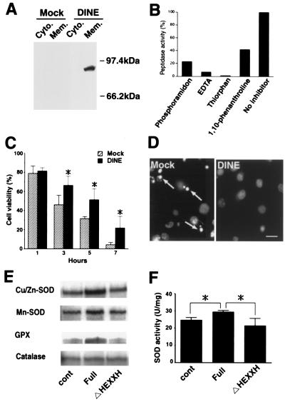
Characterization of DINE protein. (A) Western blotting using cytosol (Cyto.) and membrane (Mem.) fractions collected from COS-7 cells, which were transfected with either full-length DINE or mock. (B) Proteolytic activity of recombinant DINE. For inhibition experiments, typical metalloprotease inhibitors were added before incubation with substrate. (C) C2-ceramide-induced apoptosis in COS cells was significantly inhibited by the expression of DINE. Cell survival assay using trypan blue was carried out. Hatched bar, vehicle-plasmid expressing cells; closed bars, DINE-expressing cells. Bars represent mean +/− SD; *, P < 0.05 (ANOVA test). (D) Apoptotic nuclei (arrows) were identified by staining with Hoecst 33528 dye 5 h after exposure to C2-ceramide. (Bar = 20 μm.) (E) RNase protection assay showed that the proteolytic activity of DINE promotes Cu/Zn-SOD, Mn-SOD, and GPX expression, but not catalase at 1 h after C2-ceramide exposure. Cont, vehicle-plasmid expressing cells; Full, full-length DINE-expressing cells; ΔHEXXH, mutated DINE, lacking zinc-binding domain, expressing cells. (F) SOD activity induced by proteolytic activity of DINE. Bars represent means +/− SD; *, P < 0.05 (ANOVA test).

