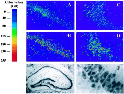
Figure 1.
GPx1-like immunoreactivity in areas CA1 and CA3 of hippocampal sections prepared from Tg-GPx mice. Pseudocolor representations of immunoreactivity in areas CA1 (A and B) and CA3 (C and D) of the hippocampus of non-Tg (A and C) and Tg-MT-GPx-6 (+/+) (B and D) mice. Photomicrographs A and C show GPx-like immunostaining in areas CA1 and CA3 of non-Tg mice, respectively. Note the more intense staining (red shift) in the cell body layer in both the CA1 and CA3 regions in Tg mouse as compared with non-Tg mouse. (E) GPx-like staining in the whole hippocampal region of the Tg mouse (low magnification; ×20). (F) High-magnification (×400) picture of a CA1 segment of hippocampal section shown in E, showing intense staining in the pyramidal cells.

