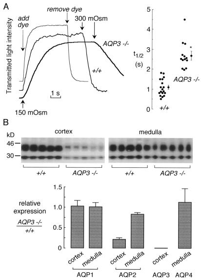
Figure 4.
Effect of AQP3 deletion on collecting-duct water permeability and expression of renal aquaporins. (A) Osmotic water permeability in basolateral membrane of cortical collecting ducts. (A Left) Representative time courses of collecting-duct cell volume (genotype indicated). Perfusion with hypoosmolar solution (150 mosM) produced cell swelling and increased transmitted optical signal. Perfusion exchange time (labeled “add dye” and “remove dye”) was determined by absorbance of the dye Evans blue. (A Right) Averaged swelling rates for indicated mouse genotypes (SEM; *, P < 0.001). (B) Effect of AQP3 deletion on expression of renal aquaporin proteins. (B Upper) Immunoblot analysis of AQP2 in membranes from renal cortex and medulla from six wild-type and six AQP3-knockout mice. (B Lower) Quantitative densitometry of immunoblot data for indicated aquaporins.

