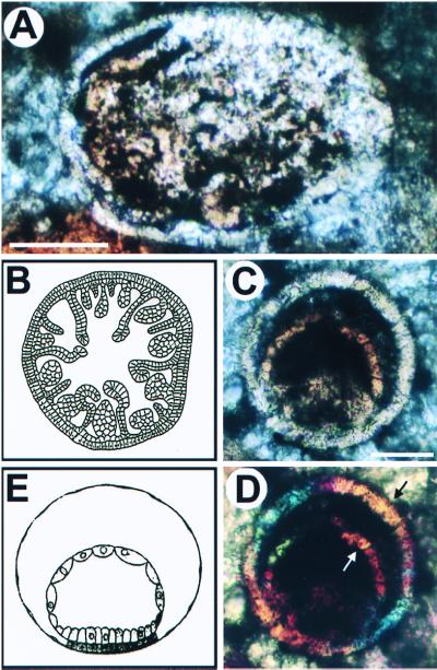
Figure 2.
Putative cnidarian embryos and larvae. (A) Oblique section of a possible fossil anthozoan planula. (B) Schematic view of a transverse section of the late planula of the anthozoan Euphyllia rugosa. The larval stage represented in A and B is constituted of an outer monocellular layer, the ectoderm, within which is an inner endodermal layer with various mesenteric folds and immature septa. This complicated bilayered structure is typical of anthozoan late planula larvae. Note the individual cells visible in the ectodermal layer at lower left in A, where it has separated from the endodermal layer. (Scale bar, 100 μm.) (C and D) Putative fossil gastrula of hydrozoan medusa; (C) Bright field; (D) Polarized light. Under polarized light (D), both layers show the same crystal orientation at arrows, as indicated by the same colors. The modern hydrozoan embryo shown in E is Liriope mucronata. B is from Chevalier (47); E from Campbell (48). (Scale bar in C is 50 μm.)

