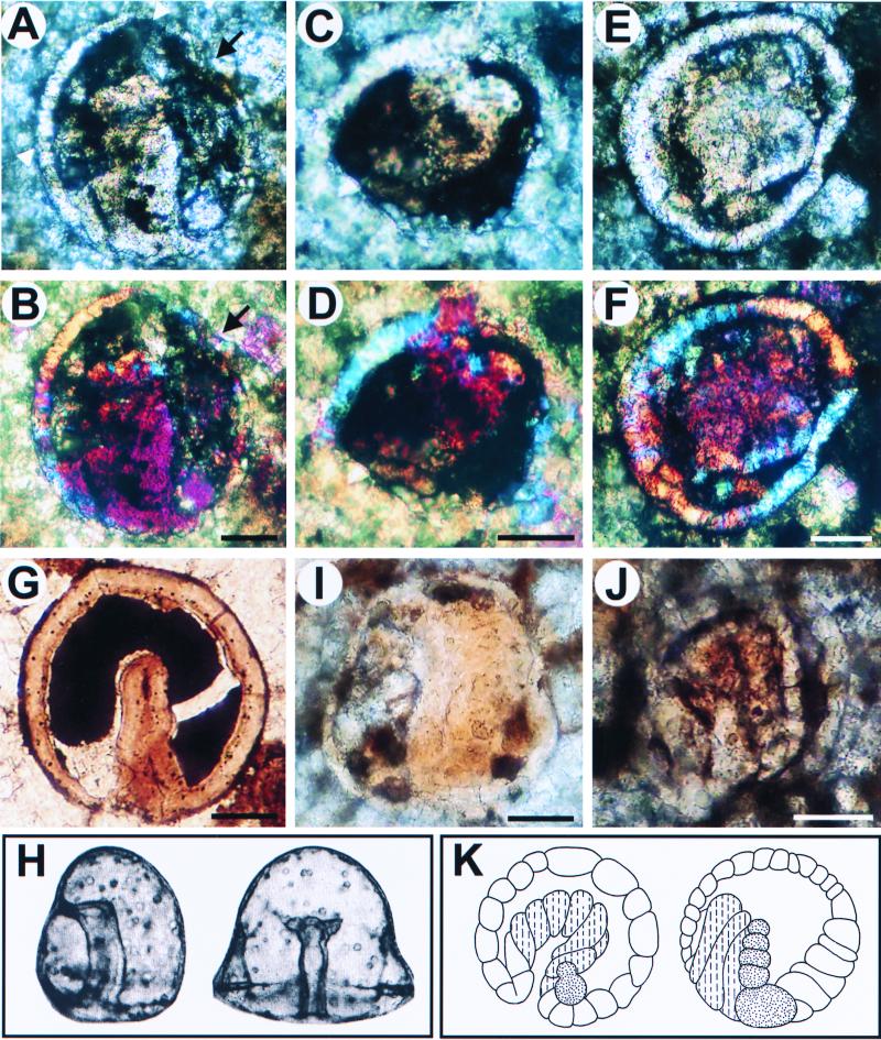
Figure 3.
Putative fossil embryos that resemble bilaterian gastrulae. (A–G) Fossils resembling deuterostome embryos; (H) Modern example (gastrulae of the sea urchin Mespilia globulus, ref. 49) In A, C, and E, the archenteron is bent to one side, and in A and C displays bilobed outpocketings; (A) The nearer ectodermal layer is thicker compared with the opposite one (possible oral and aboral ectoderms, respectively; compare H). (C) A section in the plane indicated by the small arrowheads in A. (B and D) Polarized light microscope images, showing that the cells comprising the outpocketings are differently oriented, as they appear in different colors from those constituting the walls of the gut. In A, part of the outer wall is deformed (arrow) by a crystal grain visible in B (light pink). (G), Another specimen displaying invaginating archenteron at early midgastrula stage. (H) Modern sea urchin gastrulae (49). (I and J), Fossils resembling modern spiralian gastrulae; (K) Modern polychaete embryos in which the dashed lines indicate yolky endoderm cells and dots represent mesoderm cells (Eupomatus, left; Scoloplos, right, redrawn from Anderson, ref. 50). In the fossils I and J, the archenteron is thick-walled (cf. cross section in C), and in J all of the cells in the embryo, including the ectodermal wall, are conspicuously larger relative to the size of the embryo. Note also the column of cells along the archenteron in J. (Scale bars represent 50 μm.)

