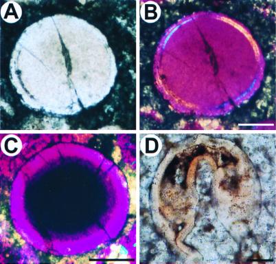
Figure 4.
Oncoids and an undefined embryo, larva, or adult form. Bright-field (A) and polarized (B) light images of an oncoid found in one of the thin sections. The inner mass consists of a unique homogeneous crystal, as shown by its uniform color in polarized light, surrounded by a laminar structure. Note the crack in the center. The two outside thin layers are uniform, and they do not delimit any cavity. (C) A smaller oncoid; note the uniform circumferential wall, which in polarized light is the same color throughout. (D) Unidentified biological form. There is a large blastocoel and a gut of some kind. The top of the fossil embryo is slightly deformed, suggesting that this structure was soft. (Scale bars are 50 μm.)

