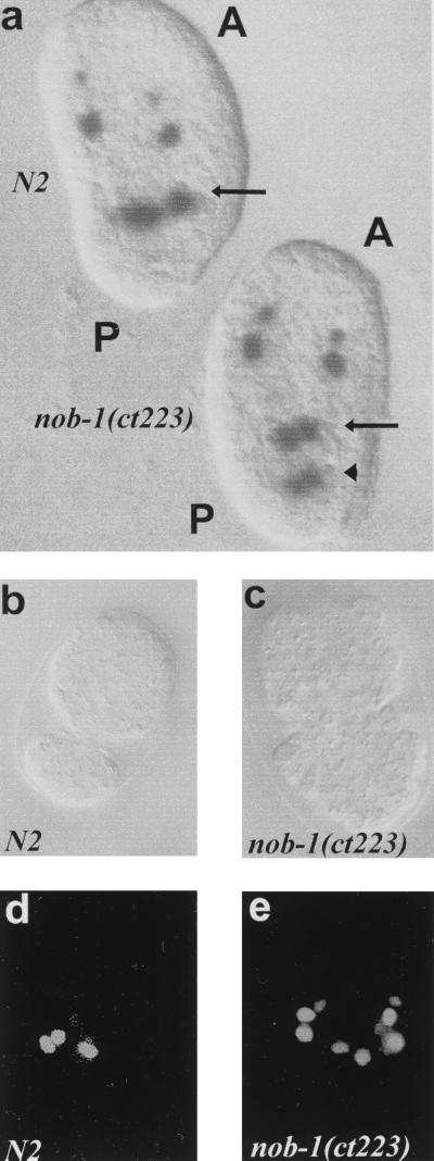
Figure 4.
Posterior-to-anterior fate transformations in nob-1(ct223) embryos. (a–c) Nomarski images with anterior (A) toward the top and posterior (P) toward the bottom. (d and e) Epifluorescence images of b and c, respectively. In a, pal-1∷lacZ expression is shown in a wild-type (upper) and a nob-1(ct223) (lower) embryo. The arrows point to the two mid-region gut cells that express the reporter in both wild-type and mutant embryos; the arrowhead indicates the ectopic expression in two more posterior gut cells of the nob-1(ct223) embryo. Analysis of several such embryos with a pal-1∷GFP as well as the pal-1∷lacZ reporter identified the ectopically expressing cells as those of the int8/9 intestinal segment. The four additional anterior expressing cells are myoblasts not affected in nob-1(ct223). (b and c) Ventral views of wild-type and nob-1(ct223) embryos, respectively, during early morphogenesis when the embryo is enclosing. (d and e) Expression of the ceh-13∷GFP reporter in mid-ventral neurons of the same two embryos, respectively. Lineaging shows that the ectopically expressing cells in such Nob embryos are derived from more posterior lineages than the normally expressing cells. (This anteriorization is not evident from the figure, because at this stage, the Nob embryo is already morphologically abnormal.)

