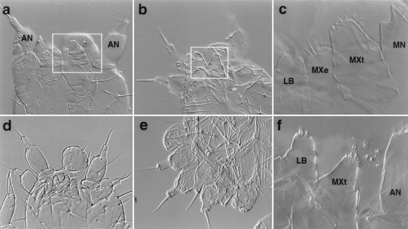
Figure 3.
Wild-type and mutant first instar larval cuticles. (a) Ventral view of a wild-type cuticle. (b) Ventral view of TcDfd1 homozygote. The terminal seta of the homeotic antenna on the right is missing. (c) The boxed region in a photographed at higher magnification. (d) Ventral view of TcDfd1/Df(HOMC). The homeotic antennae on the mandibular segment are fully transformed. (e) Lateral view of TcDfd2/Df(HOMC). Note both normal and homoetic antennae. (f) The boxed region in b photographed at higher magnification. The maxillary endite is missing, and the maxillary telopodite is reduced in size. The homeotic antenna is out of the plane of focus. AN, antennal; LB, labium; MXe, maxillary endite; MXt, maxillary telopodite; MN, mandible; LR, labrum. Lower magnification = ×200; higher magnification = ×400.

