Fig. 3.

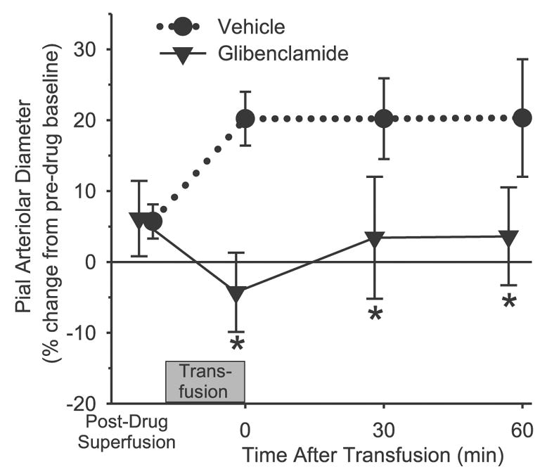
Change in pial arteriolar diameter after superfusion of the cranial window with 0.1% DMSO vehicle (n = 6) or 10 μM glibenclamide (n = 6), and at 0, 30, and 60 min after completion of an exchange transfusion of the albumin solution. Values are means ± SD and are expressed as a percentage of the baseline diameter before vehicle or drug superfusion of the window. *P < 0.05 from vehicle group.
