
Figure 4B.
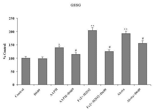
Shows the increased level of GSSG in brain mitochondria isolated from saline-injected gerbils and subsequently treated with AAPH, Fe2+/H2O2 or Aβ (1-42) as compared to GSSG levels in brain mitochondria isolated from saline-injected gerbils but not subjected to treatment of any oxidant. The reduction in GSSG level shows in brain mitochondria isolated from gerbils previously injected i.p. with D609 1 h before sacrifice and treated with AAPH, Fe2+/H2O2 or Aβ (1-42) compared to GSSG levels in brain mitochondria isolated from saline-treated gerbil and then treated with oxidants. *p<0.01 and **p<0.001 compared to control, # p<0.01 and ## p<0.01 compared to oxidant treatment. The data are presented as mean ± SEM expressed as percentage of control (n=6).