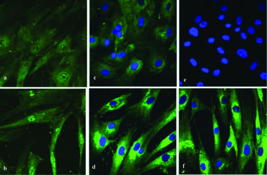
Figure 1.
Confocal scanning laser microscopic analysis of human osteoblastic cells after oxandrolone treatment. (a) Confocal scanning laser microscopy depicting AR nuclear fluorescence of human osteoblastic cells not stimulated with oxandrolone, (b) confocal scanning laser microscopy depicting the increased AR nuclear fluorescence of human osteoblastic cells stimulated with oxandrolone 30 μg/mL, (c) confocal scanning laser microscopy depicting cytoplasmic fluorescence of type I collagen in human osteoblastic cells not stimulated with oxandrolone, (d) confocal scanning laser microscopy depicting increased cytoplasmic fluorescence of type I collagen in human osteoblastic cells stimulated with oxandrolone 30 μg/mL, (e) internal negative control for primary antibodies of AR and type I collagen, (f) confocal scanning laser microscopy depicting increased cytoplasmic fluorescence of type I collagen in human osteoblastic cells stimulated with oxandrolone 15 μg/mL. Compare to Figures 1c and 1d.

