
Figure 5. Migration and migratory paths of NPCs in the adult mouse spinal cord in response to motor neuron degeneration in ALS-like mice.
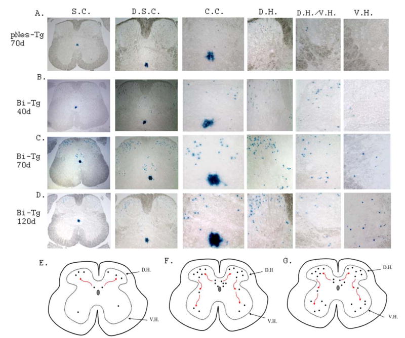
The migration of NPCs characterized by LacZ staining from the ependymal zone surrounding central canal region to the dorsal direction, and subsequently to ventral direction in control pNes-Tg mice (A: at 70 days of age), in ALS-like Bi-Tg mice during disease free (B: at 40 days of age), disease onset (C: at 70 days of age) and disease progression (D: at 120 days of age). The migratory paths of NPCs at different stages were presented in E (for normal and clinical disease free stages), F (for disease onset) and G (for disease progression) respectively.