
Figure 6. Identification and determination of the source of increased NPCs in the dorsal and ventral horn regions of the adult Bi-Tg mouse spinal cords in response to motor neuron degeneration in ALS-like mice.
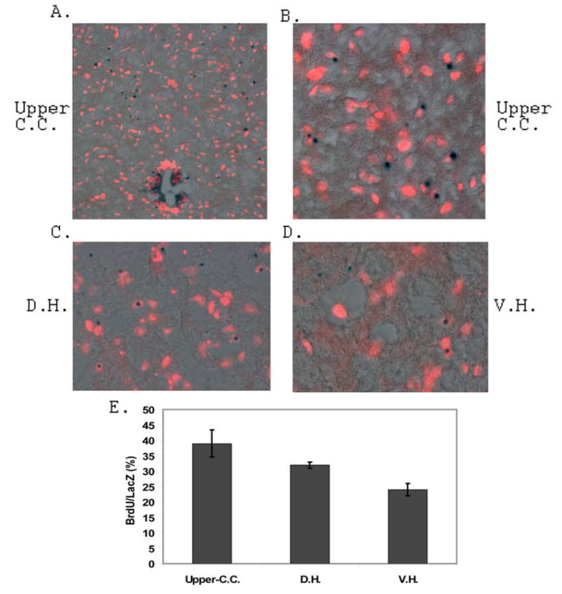
Analysis of the source of increased NPCs in the dorsal and ventral horn regions of the ALS-like mouse spinal cords was carried out by 25 days of BrdU pulse labeling and 5 days of chasing experiments. The BrdU labeled and LacZ stained NPCs in the upper region of central canal, dorsal horn and ventral horn regions of the 115 days of age Bi-Tg mice were shown in A, B, C and D respectively. The ratio of BrdU labeled LacZ positive cells in the 115 days of age Bi-Tg mice were shown in E (3 sections/mouse, n=3 mice).