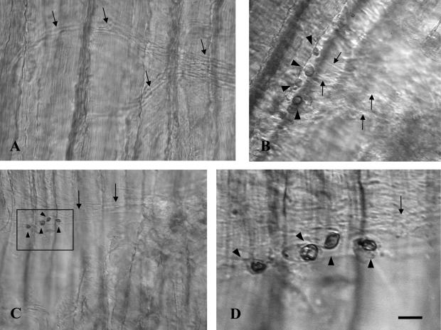
Fig. 2.
In situ hybridization in adult rat soleus using LPA3 sense cRNA probe as a negative control (A) and LPA3 antisense cRNA probe (B–D). Cell bodies of terminal Schwann cells on the rat soleus were stained (black arrowheads in B, C, D). Unstained axons were visualized under a differential interference contrast microscope (black arrows). D is a higher magnification image of the area indicated by a rectangle in C. Longitudinal and horizontal stripes were muscle fibers. Bar=50 µm (A), 20 µm (B), 40 µm (C), and 10 µm (D).

