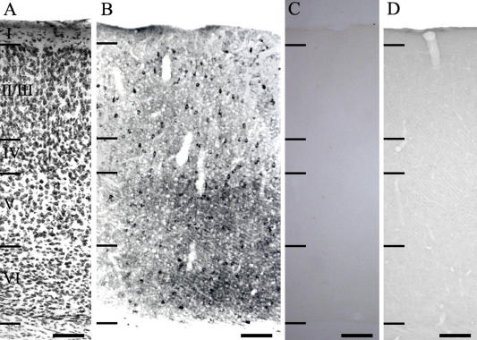
Fig. 1.
Low power photomicrographs of the distribution of GluR1-IR neurons in the hamster visual cortex. (A) Thionin-stained section shows the cortical lamination. (B) Anti-GluR1-IR neurons. (C, D) Control sections used to show the specificity of GluR1 antibody. In C, section was incubated without the addition of the primary antibody. In D, the GluR1 antibody was preabsorbed with antigen prior to tissue incubation. I–VI: cortical layers. Bar=100 µm.

