Fig. 5.
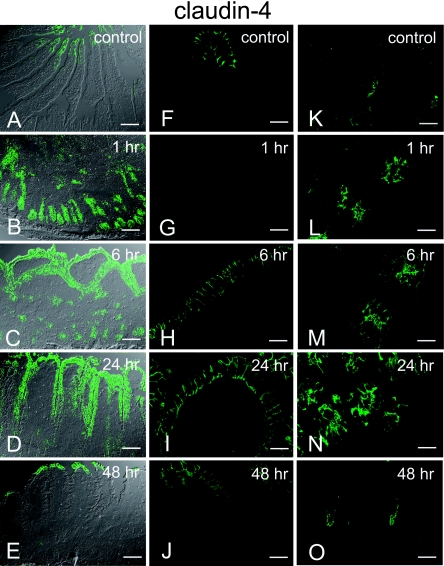
Images of immunohistochemical staining for claudin-4 obtained from a single confocal plane acquired with/without DIC imaging. Lower magnification images: (A)–(E). High-magnification images of the villi: (F)–(J). High-magnification images of the crypt: (K)–(O). Control images: (A), (F), (K). Claudin-4 expression is restricted to the tips of the villi (A, F) and to the extreme basal part of the crypt preferentially limited to the apical side of the cell (K). At 1 hr post-reperfusion (B, G, L), no expression on the surface is observed because villi are denuded (G). Claudin-4 expression remains at the crypt base with apical and lateral expression (L). At 6 hr post-reperfusion (C, H, M), strong expression of claudin-4 is observed at the surface of the epithelium with lateral expression (H). The strong expression at the crypt persists (M). At 24 hr post-reperfusion (D, I, N), claudin-4 is still extensively expressed from the tips to the villi with a diminishing gradient (D). The lateral expression in the villi (I), and apical and lateral expression in the crypt (N) are still observed. At 48 hr post-reperfusion (E, J, O), expression of claudin-4 is restricted to the tips of the villi (E, J) and to the lower base of the crypt (O), which is similar to the expression pattern observed in the control sample. Bars=20 µm (A–E), 60 µm (F–O).

