
FIGURE 2.
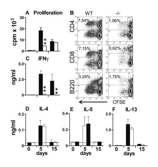
Ag-specific in vitro lymphocyte proliferation and IFN-γ production by cells from the sdLN are dependent on CD154. Cells from WT (■) and CD154−/− mice (□) obtained at days 0, 5, and 15 after parasite exposure were stimulated in vitro in the presence of exogenous SSAP. A, Total proliferation was measured by [3H]thymidine incorporation, and B, CD4+, CD8+, and B220+ division at day 5 was measured following CFSE labeling (values are percentage of cells that have divided in culture). C, E, and F, Production by sdLN cells of IFN-γ, IL-5, and IL-13 in response to SSAP stimulation, and D, IL-4 in response to anti-CD3 mAb. Dotted lines represent the minimum detection level. Data shown are cell proliferation or cytokine production in the presence of Ag or anti-CD3 mAb. In the absence of stimulus, proliferation was <1000 cpm (data not shown), and cytokine production was at the minimum level of detection. Significant differences are shown between WT and CD154−/− mice.