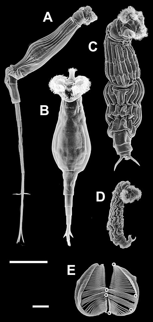
Figure 1. SEM Pictures of Some Species of the Genus Rotaria .
(A) R. neptunia, lateral view; (B) R. macrura, ventral view; (C) R. tardigrada, dorsal view; (D) R. sordida, lateral view; and (E) trophi of R. tardigrada with open circles showing the location of landmarks used for the shape analysis. Scale bars: 100 μm for animals, 10 μm for trophi.

