Figure 9.
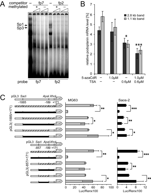
The role of PDPN promoter methylation in MG63 cells. A) EMSA competition experiments were performed using cold methylated and unmethylated fp7 (site Sp.4) and fp2 (site Sp.2) probe as competitors. Competitors were used at 50-fold excess at the small end of the triangle and at 100-fold excess at its large end. B) Densitometric quantification of both podoplanin mRNA transcripts isolated from MG63 cells which had been treated with 5-azaCdR and TSA alone or in combination. The abundance of podoplanin transcripts was normalized to GAPDH transcripts in the same samples. The data are given as the mean ± S.D. of triplicate samples of the experiment repeated twice. The statistical analysis denotes the significant difference between non-treated MG63 cells and cells treated with 5-azaCdR or/and TSA. *, p < 0.05, ***, p < 0.001. C) Whole pGL3-constructs or selected inserts thereof were methylated or mock-methylated in vitro and transfected into MG63 and Saos-2 cells. Results are given as luciferase activity normalized to cotransfected pRL-TK activity. The experiment was repeated three times independently in triplicates. The statistical analysis denotes the significant difference between mock methylated (hatched) and respective methylated (black) constructs. *, p < 0.05, **, p < 0.01, ***, p < 0.001.

