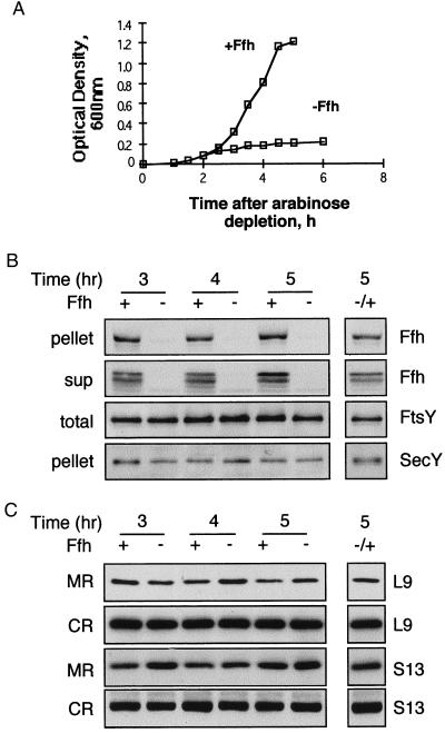
Figure 4.
Effect of Ffh depletion on the cellular distribution of ribosomes. (A) Growth of E. coli WAM113 with or without arabinose was monitored by measuring the optical density of the cultures every 30 min. Samples were taken at certain time points during the growth, fractionated as described for Fig. 1, and analyzed by Western blotting as shown in B (ultracentrifugation of cell extracts) and C (flotation assay). (B and C) In both panels, the separated lane on the right represents samples from depleted cultures that were induced with arabinose after 4 hr of depletion and harvested 1 hr later. (C) Extracts were prepared from WAM113 cells that were grown with or without arabinose (A). Pellets (10 μg of proteins) were probed with anti-Ffh and anti-SecY antibodies; supernatants (sup; 20 μg of proteins) were probed with anti-Ffh antibodies; and total extracts (total; 10 μg of proteins) were probed with anti-FtsY antibodies. (C) After flotation, identical aliquots from the interface fractions (membrane ribosomes, MR) and the bottom fractions (cytoplasmic ribosomes, CR) were probed with antibodies against the ribosomal proteins L9 and S13.

